An echocardiogram can be used to determine the source and extent of cardiac pathology. An echocardiogram can be useful as a baseline when a murmur is first heard or prior to any surgical procedures to assess for anesthetic risk. In patients with clinical signs of coughing or difficulty breathing, we can look for evidence of primary heart disease as a cause of the clinical signs and to rule in/out the possibility of heart failure. Thoracic radiographs in conjunction with an echocardiogram can be helpful as they will better evaluate the lungs.
Echocardiograms are performed utilizing color flow, pulse wave and continuous wave doppler. Exams include standard right parasternal and left apical imaging with stills and cine loop images. Formal cardiologist reporting will include assessment and treatment recommendations. The consulting cardiologist will also help you decide when referral is necessary if it is deemed that the patient would benefit from management by an attending cardiologist such as in patients with congenital abnormalities. The consulting cardiologist will be available for questions by phone or email.
A blood pressure measurement (PetMap) and 6 lead EKG may also be useful and is an additional service offered by Mettasound.
Indications and assessments:
Heart murmurs
Enlarged heart on x-rays
Coughing
Difficulty breathing
Collapse/fainting
Abnormal heart rhythms
Hypertension (elevated blood pressure)
Pre-anesthetic screen
Pleural effusion or pericardial effusion (fluid around the lungs or heart)
Heart based masses or right atrial masses/cancer screen
Masses in the chest
Diaphragmatic hernia
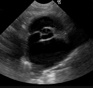
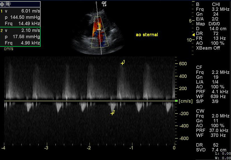
Click on image for larger view.
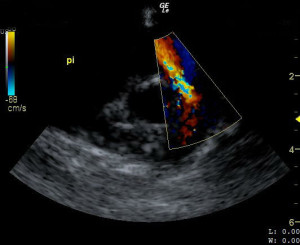
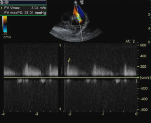
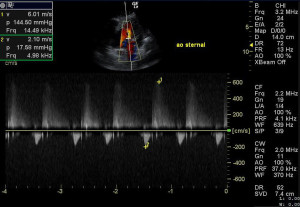
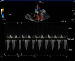
Pulmonary insufficiency in a dog with HW diseaseCW doppler in same patient showing PI velocity of 3 m/s, pulmonic insufficiency velocity greater than 2.2 m/s is generally considered to be diagnostic for diastolic pulmonary hypertension.Aortic insufficiency in a dog with systemic hypertensionMitral Valvular insufficiencyLeft atrial enlargement in a cat with HCM